_PNG.png)
Spine Anatomy 101: What Does "L4-L5" or "C5-C6" Even Mean?

Just got your MRI results back and feeling confused? Dr. Rafath Baig breaks down spine anatomy so you can understand what terms like "L4-L5" and "C5-C6" actually mean for your back pain.
Getting an MRI report back can feel like trying to read a foreign language. You are suddenly staring at phrases like "L4-L5 disc herniation" or "C5-C6 stenosis," left to wonder what those random letters and numbers actually mean for your body and your future.
When you are in pain, the anxiety of not understanding your own medical documents only makes the situation more stressful. At Desert Spine and Scoliosis Center, Dr. Rafath Baig believes that no patient should ever be handed a diagnosis they don't understand. If you are experiencing back or neck pain, the first step to finding relief is understanding exactly where it is coming from.
Here is a quick, plain-English guide to your spine's anatomy, how to read those MRI results, and why becoming a "mini spine expert" is the key to making the best decisions for your health.
Decoding the Letters and Numbers

Your spine is a column of individual bones called vertebrae, stacked on top of one another. To make it easy to identify exactly where a problem is, doctors divide the spine into different regions (the letters) and number the bones from top to bottom (the numbers).
Here is the cheat sheet for how your spine is organized:
Cervical Spine (C1 to C7): This is your neck. It starts right at the base of your skull (the occiput) and goes down to the top of your shoulders. These seven vertebrae are responsible for supporting the weight of your head and allowing you to turn, nod, and tilt your neck.
Thoracic Spine (T1 to T12): This is your mid-back, which connects to your rib cage. Because these vertebrae are attached to your ribs, this section of the spine is highly rigid and designed to protect your vital internal organs. Thanks to this limited mobility, it is relatively rare to suffer a herniated disc in the thoracic spine.
Lumbar Spine (L1 to L5): This is your lower back. Because it carries the vast majority of your body's weight and handles a lot of movement, the vertebrae here are the largest. The lumbar spine is the core of your mobility, allowing you to bend, lift, and twist.
Sacrum (S1 to S5): This is the very base of your spine, sitting right between your hip bones. Unlike the rest of your spine, these five bones naturally fuse together as you enter adulthood to form a solid triangle that connects your spine to your pelvis. Below the sacrum sits the coccyx, commonly known as the tailbone.
So, what about the discs? The shock-absorbing discs in your back sit between these bones. Therefore, they don't have their own individual numbers. Instead, they are named for the bones they connect. For example, if you have a herniated disc located between your fourth and fifth lumbar vertebrae, your MRI will read "L4-L5."
The Anatomy of a Spinal Disc
To fully grasp your MRI report, it helps to understand how these intervertebral discs are built. You can picture a healthy spinal disc much like a jelly donut.

The Annulus Fibrosus: This is the tough, fibrous outer ring of the disc (the dough of the donut). It provides structure and protects the inside.
The Nucleus Pulposus: This is the soft, gel-like center of the disc (the jelly). It provides the crucial shock-absorption and hydration your spine needs to bend and flex without the bones grinding against one another.
When your MRI report mentions a "disc tear," "bulge," or "herniation," it is referring to damage occurring to this jelly donut structure.
The Two Major Trouble Zones
While your spine is a long, complex structure, the vast majority of patients seeking treatment share something in common. Roughly 90% of all spine issues happen in just two areas: the Cervical (neck) and the Lumbar (lower back) spine.
Because these areas are highly mobile and bear a lot of stress, they are much more prone to wear-and-tear, herniations, and degenerative changes than the rigid Thoracic spine. Understanding which zone your issue is in helps explain your symptoms:
Cervical Issues (like C5-C6): Often present as pain starting in the neck that radiates down into the shoulder, arm, or fingers. The C5-C6 and C6-C7 levels are the most commonly injured areas in the neck due to poor posture (such as "tech neck" from looking down at phones) or whiplash from car accidents. Damage here can cause cervical radiculopathy, leading to numbness, tingling, or a weak grip in your hands.
Lumbar Issues (like L4-L5): Often present as lower back pain, or "sciatica"—pain that shoots from the lower back down through the buttocks and into the legs. The L4-L5 and L5-S1 segments take the brunt of your body's mechanical stress. They act as the fulcrum when you bend over to lift something heavy. Because they endure so much pressure, they are the most frequent sites for severe disc herniations and degenerative disc disease.
Translating Other Common MRI Buzzwords
In addition to the letters and numbers, your MRI report likely contains a few other clinical terms. Here is a quick translation guide for the most common diagnoses:
Bulging Disc: The tough outer layer of the disc (annulus fibrosus) is intact, but the disc has flattened and is bulging outward, much like a hamburger patty that is too big for its bun.
Herniated Disc: The tough outer layer has actually torn, and the soft jelly inside (nucleus pulposus) is leaking out. If this leaking jelly presses against a nearby spinal nerve, it causes intense, radiating pain.
Spinal Stenosis: "Stenosis" simply means "narrowing." Over time, due to bone spurs, thickened ligaments, or collapsing discs, the open spaces within your spine can narrow. This compresses the spinal cord or the branching nerves, causing pain, cramping, and weakness when walking.
Degenerative Disc Disease (DDD): This is not a true "disease," but rather the natural wear-and-tear of the spine over time. As we age, our discs lose hydration, shrink, and become less effective at absorbing shock.
Why You Need to Become a "Mini Spine Expert"
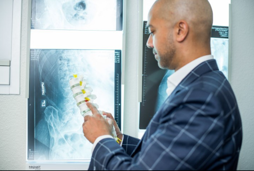
It can be incredibly daunting to sit in a doctor's office and simply be told, "You need surgery." It is even more frustrating when the doctor does not take the time to explain why.
Dr. Baig's Philosophy: "It is important for the patient to understand what we are looking at, rather than simply telling them they need surgery... I always believe that the most informed patient will always make the best decision as to what the course of action will be next."
When you come into the office, the goal isn't just to treat you; it is to educate you. By walking through your MRI slice by slice, pointing to the spinal models, and explaining the exact anatomy causing your pain, you become a momentary "mini spine expert" of your own body.
When you know exactly what is going on with your spine—whether it's a disc bulge, stenosis, or a cyst—you take the power back. This knowledge empowers you to understand why conservative treatments (like physical therapy or injections) might work, or why an ultra-minimally invasive surgery or artificial disc replacement might be the best path forward to protect your mobility.
Ready to decode your MRI?
If you have an MRI report that you need help understanding, or if you are dealing with chronic back or neck pain that is getting in the way of simple daily activities, give us a call.
Contact Desert Spine and Scoliosis Center today to schedule a comprehensive consultation. Let our team help you translate your pain into a clear, actionable plan to get you back to real living.



Comments